Orbital Roof Reconstruction

Ali Badkoobehi Presenting Www Dralibadkoobehi Com Badkoobehi Oralandmaxillofacialsurgery Dentists Facial Reconstruction Surgery Facial Bones Surgery

This Page Is For The Anthropology Section Of The Course Which Took Approximately 3 Days Of 40 Minute Periods Forensics Forensic Facial Reconstruction Skull

Cmf Case Study 1 Cranioplasty And Orbital Roof Reconstruction 3d Lifeprints
Orbital Reconstruction For Orbit Orbital Floor Fracture

Neurofibromatosis Type 1 Orbital Manifestations Radiology Case Radiopaedia Org Dysplasia Of The Neurofibromatosis Type 1 Manifestation Radiology Imaging

Pdf Early Reconstruction Of Orbital Roof Fractures Clinical Features And Treatment Outcomes Semantic Scholar
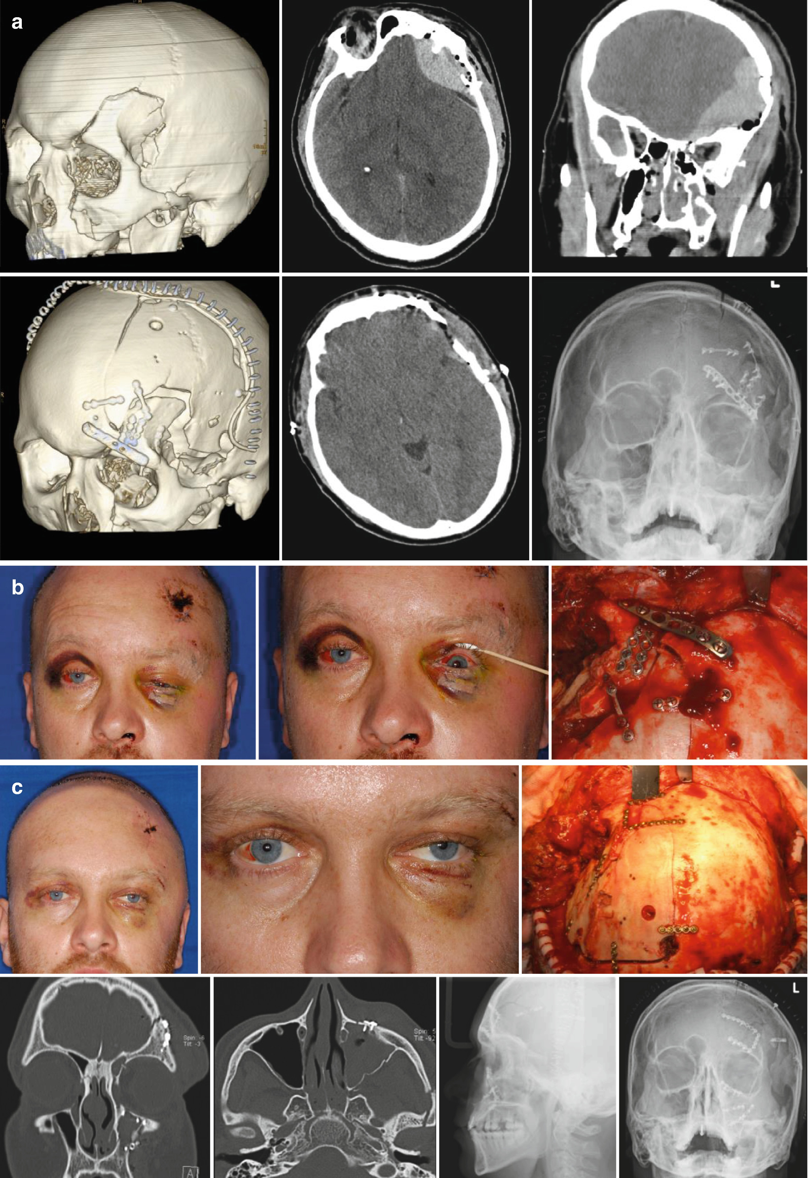
The anterior fossa is exposed extradurally with visualization of the anterior two thirds of the orbital roof.
Orbital roof reconstruction.
Secondary Reconstruction Of Posttraumatic Orbital Deformities World Renowned Bespoke Cosmetic Plastic Surgeon Boston Dr Michael Yaremchuk

Figure 4 From Reconstruction Of Orbital Roof Fracture Using Titanium Mesh Case Report And Review Of Literature Semantic Scholar

Frontal Sinus Reconstruction Surgery Part 1 Medical Illustration

Burst Fracture Of Lumbar Spine Sagittal Reconstruction Of Ct Of The Lumbar Spine Demonstrates A Comminuted Vertical Burst Fracture Through The Body Of L1 Whit

View Image

Google Image Result For Https Www Ancient Eu Img R P 500x600 1440 Jpg V 1485680505 Aztec Civilization Aztec City Aztec Capital

Wmf Office On Behance Entrance Gates Design Facade Architecture Pavilion Architecture

Post Operative Ct 3d Reconstruction Showing Metallic Prosthesis Seen Download Scientific Diagram

Gammel Hellerup Gymnasium Big Architects Architecture Architect
Remarkable Facial Reconstruction Surgery Medizzy Journal

Frombork Warmia Poland Visit Poland Poland Beautiful Places

Pin On Arq

Modern Loft Building Hice

Elevated Building Foundation At Reconstruction Glendale California Aswinkr Courtesy Civil Work Building Foundation Steel Buildings Structure Design

8 Ruined Cities That Remain A Mystery To This Day Catal Huyuk Ancient Humans Ancient Cities
Orbitotomy With Orbital Roof Repositioning For Supraorbital Ridge Orbital Roof Malposition

Millions Pledged To Rebuild Notre Dame We Will Rebuild Cathedral Paris Landmarks
Open Treatment For Orbit Orbital Roof Fracture
Https Encrypted Tbn0 Gstatic Com Images Q Tbn 3aand9gcr9ebtrfmvh1rzufodair3y6kj1cx4f0nvjppqkixoyldrrk5jk Usqp Cau

Fire Search Results Ancient Archives En 2020 Archeologie Fresque Souterrain

A New 100 M 500 Mbps Underwater Optical Wireless Communication System Advances In Engineering Communication System Communication Information And Communications Technology

1 698 Likes 22 Comments Trades Directory Tradectory On Instagram This Roof Opening With Balcony Is House Design Architecture House Architecture Design

Fd Lymphatic Intravasation This Hsg Shows A Streaky And Mesh Like Collection Of Contrast Material In The Right Side Of Th Lymphatic Material Radiology

Copper Beaten Skull Also Known As Beaten Brass Skull Refers To The Prominence Of Convolutional Markings Gyral Impressions On The Inner Table Of The Skull Tip
Radioogle Ent Right Orbital Roof Reconstruction

Base And Top In Architecture Facade Design Facade Architecture Architecture Plan
Surgical Strategy In Complex Craniofacial Trauma Care The Expert S Experience And Suggestions Springerlink

Mvrdv Completes The Stairs A Giant Scaffolding Staircase In Rotterdam City Centre

Ryobi Corner Cat Sander 1 2 A Ryobi Best Random Orbital Sander Finishing Sander

Radiology Rounds Radiology Rounds Medical Specialties Medical Training Radiology

The Pinch Zhaotong 2014 John Lin Olivier Ottevaere Centro Comunitario Arquitectura Biblioteca Arquitectura
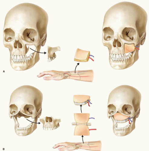
Reconstruction Of The Maxilla And Skull Base Plastic Surgery Key

The Proposed New Stadium For The Newly Acquired Nfl Team The Los Angeles Rams Will Be Delayed Until 2020 V Nfl Stadiums San Diego Chargers Football Stadiums
Hie Multimedia Head And Face Reconstruction

32 Essential Home Improvements To Make When You Win The Lottery Rural House Architecture House Design

Proposed Four Films Printing Factory F M Offices Printing Skyscrapercity Factory Architecture Steel Structure Buildings Light Art Installation

A Craniofacial Initial Ct Case 3 3d Reconstruction Panfacial Download Scientific Diagram

Pin On Radiology

Pin On X Ray Abdomen

Systematic Analysis Of Skull Base Cranial Vault Fractures A Download Scientific Diagram

Novartis Campus In Basel By Frank Gehry Serge Ferrari Low Emissivity Solar Protection Screens Amazing Buildings Architecture Design Unique Architecture

The Red Arrows Indicate The Location Of The Closed Coronal Sutures Note The Increased Height And Width Of The Skull Cleft Lip And Palate Cleft Lip Rhinoplasty

Pin Em Videos

Le Corbusier Five Points Of Architecture As Seen In The Villa Savoye Bauhaus Architecture Le Corbusier Le Corbusier Architecture
Source : pinterest.com
